
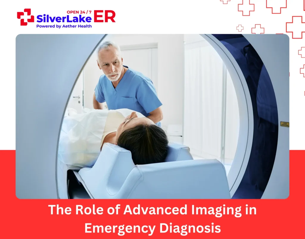
A sudden injury or unexplained pain creates a cascade of questions. You need answers quickly, not tomorrow after a lengthy referral process. Advanced imaging technology reveals what’s happening inside your body during these critical moments.
Our ER physicians rely on CT scans, digital X-rays, and ultrasounds to see beyond surface symptoms. These powerful diagnostic tools help distinguish between conditions that present similarly but require drastically different treatments.
At our emergency facilities in Silverlake, we provide immediate access to advanced imaging technology with onsite radiologists who interpret results quickly. Our integrated approach means you receive accurate diagnosis and appropriate treatment without unnecessary delays.
Importance of Advanced Imaging in Emergency Care
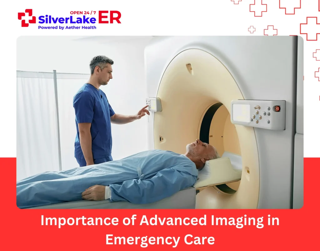
Emergency departments function with speed and accuracy, as patients frequently come in with life-threatening conditions. Standard physical examinations and medical history evaluations offer important insights, but advanced imaging provides a more thorough understanding of internal injuries and illnesses.
Diagnostic imaging is particularly critical in cases:
- Trauma and injuries – Identifying fractures, internal bleeding, and organ damage
- Neurological crises – Detecting strokes, brain hemorrhages, and spinal cord injuries
- Cardiac incidents – Assessing heart function and excluding life-threatening conditions
- Pulmonary concerns – Diagnosing pulmonary embolisms, collapsed lungs, and pneumonia
- Abdominal pain – Recognizing conditions like appendicitis, gallstones, or internal bleeding
By incorporating advanced imaging into emergency treatment, Silver Lake ER improves diagnostic precision for timely and life-saving treatment.
Computed Tomography (CT) Scans
CT scans stand as the cornerstone of emergency imaging because they create detailed cross-sectional images in minutes. This technology captures multiple X-ray images from different angles then digitally reconstructs them to show bones, organs, soft tissues, and blood vessels in exceptional detail. In emergencies, CT scans deliver the clarity physicians need to make life-saving decisions.
When Are CT Scans Used?
- Head injuries and strokes – A non-contrast CT scan is the preferred initial imaging option for analyzing traumatic brain injuries and strokes. It identifies bleeding in the brain (intracranial hemorrhage), skull fractures, and indications of ischemic strokes by locating areas of diminished blood flow.
- Trauma cases – In cases of blunt force trauma or accidents, a whole-body CT scan, also referred to as a “pan-scan,” helps in identifying internal bleeding, organ lacerations, fractures, and other critical injuries. The capacity to gather thorough diagnostic information in mere minutes makes CT scans a critical tool in trauma care.
- Abdominal emergencies – CT scans are commonly used to diagnose issues such as appendicitis, bowel obstructions, diverticulitis, and internal bleeding. With contrast enhancement, CT imaging offers a detailed view of abdominal organs for accurate treatment choices.
- Pulmonary embolism detection – A CT pulmonary angiogram (CTPA) is the benchmark for diagnosing pulmonary embolism, a life-threatening issue caused by blood clots in the lungs. By using contrast dye to clarify blood vessels, emergency physicians can clearly visualize any blockages and recommend appropriate treatment.
CT scans are not only swift and widely accessible but also provide critical diagnostic information. These capabilities ensure that life-threatening conditions receive prompt identification and treatment.
Digital X-Rays
Digital X-rays represent a significant advancement over traditional film radiography. They offer immediate image availability with superior detail and clarity. This technology uses electronic sensors instead of photographic film to capture images, which enables instant viewing and sharing with specialists. The speed and precision of digital X-rays make them particularly valuable in time-sensitive emergency situations.
When Are Digital X-Rays Used?
- Fracture detection – Digital X-rays remain the first-line imaging choice for suspected bone fractures. They clearly show bone alignment, fracture patterns, and joint dislocations, helping physicians determine appropriate treatment from splinting to surgical intervention.
- Chest emergencies – For patients with breathing difficulties, chest X-rays quickly identify pneumonia, collapsed lungs (pneumothorax), fluid accumulation (pleural effusion), or concerning masses. The ability to magnify and adjust digital images eases detection of subtle lung abnormalities.
- Foreign body localization – When patients have ingested or inhaled foreign objects, digital X-rays help locate these items rapidly. The technology is especially useful in identifying objects that may require immediate surgical removal.
- Joint and soft tissue assessment – Beyond bones, digital X-rays help evaluate joint spaces for arthritis, effusions, or alignment problems. Enhanced contrast capabilities also improve visualization of some soft tissue problems that aren’t visible on conventional X-rays.
Digital X-ray technology delivers significant advantages in radiation dose reduction without compromising image quality. This combination of safety, speed, and diagnostic accuracy makes digital radiography an essential emergency medicine tool.
Ultrasound in Emergency Medicine
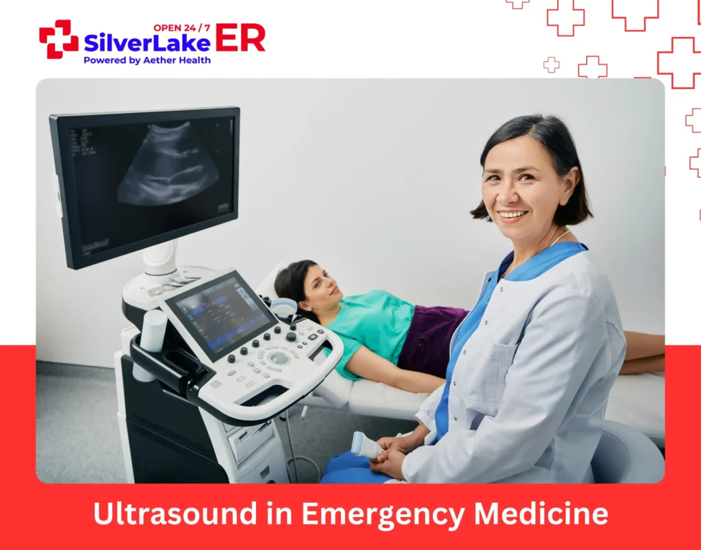
Ultrasound imaging is a non-invasive, radiation-free diagnostic method that provides real-time images of internal structures. It’s extensively used in emergency medicine for fast bedside assessments, particularly when immediate decisions are required.
When Are Ultrasounds Used?
- Cardiac emergencies – A point-of-care ultrasound (POCUS) enables emergency physicians to quickly evaluate heart function, detect pericardial effusion (fluid around the heart), and identify cardiac tamponade, a life-neutralizing condition needing urgent attention.
- Abdominal pain – Ultrasound efficiently evaluates gallstones, kidney stones, liver irregularities, and fluid accumulation in the abdominal cavity. It’s particularly beneficial for identifying conditions like cholecystitis and hydronephrosis without exposing patients to radiation.
- Pregnancy-related emergencies – In obstetric emergencies, ultrasound is critical for evaluating fetal health, diagnosing ectopic pregnancies, and detecting placental abnormalities. It supplies critical information guiding immediate obstetric care decisions.
- Internal bleeding – The FAST (Focused Assessment with Sonography for Trauma) scan is a fast ultrasound procedure conducted in trauma cases to identify free fluid in the abdominal cavity, indicating possible internal bleeding. This swift and portable assessment helps in directing resuscitation efforts.
The Role of Advanced Imaging in Trauma Case

When someone comes into the ER after a trauma, we jump right in and check for any life-threatening issues. Here at Silver Lake ER, we use a mix of CT scans, X-rays, and ultrasounds to quickly figure out what’s going on.
Situations Where Imaging Really Matters:
- Car accidents – CT scans help us examine internal injuries, brain issues, and spinal cord problems.
- Falls and fractures – X-rays are great for spotting bone breaks, but sometimes CT or MRI is needed for the trickier injuries.
- Blunt force trauma – Imaging helps us check for internal bleeding, organ damage, or brain injuries.
Fast and accurate imaging is key to making sure trauma patients get the right care without wasting any time.
Advanced imaging is a huge part of how we diagnose emergencies. Tools like CT scans, MRIs, ultrasounds, and X-rays give us important insights that guide us to act quickly and decisively.
FAQs
Why do doctors prefer CT scans?
Doctors prefer CT scans for their fast, detailed cross-sectional imaging, ideal for diagnosing strokes, internal bleeding, and complex injuries.
Why is CT better than radiography?
CT scans outperform radiography by providing 3D imaging and better tissue differentiation. It’s speed and clarity make it more effective for detecting subtle abnormalities.